
How CT Imaging Is Changing the Way We Plan Surgery
Bentonville Veterinary Specialists is proud to offer computed tomography (CT) as part of our advanced diagnostic capabilities. At BVS our 160 slice CT is state of the art and produces a better quality image than nearly every practice in the Southeast. CT allows BVS to continue offering the highest standard of care for our patients requiring advanced diagnostics.
What Makes CT Different from Conventional Radiographs?
The main benefit of CT compared to conventional radiography is its cross-sectional imaging capability. Traditional X-rays collapse a three-dimensional structure into a flat image, which means depth perception is lost. CT eliminates that limitation entirely.
With CT, our surgical team can:
- Accurately localize lesions without the guesswork of overlapping structures.
- Better define tumor borders for more complete surgical planning.
- Assess fracture orientation in three dimensions, including comminution that may be invisible on plain films.
- Evaluate spinal cord compression with far greater precision.
- Benefit from superior contrast resolution, leading to more accurate diagnosis and treatment planning.
CT may benefit patients with orthopedic, neurologic, or soft tissue surgical diseases, and is particularly useful for diagnosing abnormalities of the nasal passages, middle and inner ear, brain, spine, abdomen, lung, mediastinum, and the musculoskeletal system.
The case below illustrates exactly why this technology matters.
Case Study: Tarsal Fractures in an Eight-Month-Old German Shepherd
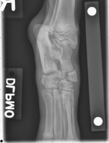
An eight-month-old German Shepherd dog presented for acute right hindlimb lameness after she was struck by a moving car. On examination, soft-tissue swelling, pain, and crepitus were present at the right tarsus.
Initial radiographs revealed:
- A type II central tarsal bone fracture.
- Fracture of the fourth tarsal bone.
- Fracture of the second metatarsal bone.
Based on the radiographic findings, repair of the tarsal fractures with lag screws was recommended as the initial surgical plan.
How CT Changed the Surgical Plan
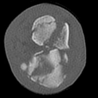
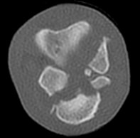
Prior to surgical correction, a CT was performed — and it revealed something the radiographs had not shown: significant comminution of both tarsal bones.
This was a critical finding. Because the fractures were:
- Highly comminuted (broken into multiple fragments).
- Involving the articular surface.
- Unable to be perfectly anatomically reduced.
...the original plan of lag screw fixation was no longer appropriate. Attempting to reconstruct these fragments would have been unlikely to restore a functional, pain-free joint.
Instead, the surgical plan was changed to a partial tarsal arthrodesis — a procedure that fuses the affected joints, eliminating the painful, unstable articulation and providing a stable, functional limb.
The Outcome
At three months following surgery, the dog's lameness had fully resolved.
This case is a clear example of how advanced imaging does more than confirm a diagnosis — it shapes the entire surgical strategy. Without CT, the comminution would not have been appreciated preoperatively, and the patient may have undergone a procedure unlikely to succeed.
Is CT Right for Your Patient?
If your pet has been referred to us or you are a veterinarian considering a referral, CT imaging is available at BVS for patients with orthopedic, neurologic, or soft tissue conditions. Our team will determine whether CT is indicated as part of the diagnostic and surgical planning workup.
Have questions? Contact us at [email protected].
Have questions about your pet's recovery?
Our surgical team is here to help — don't hesitate to reach out.
Contact Us